Imaging Biomarkers

Thomas L. Mindt, head of the program line, is a well-recognized expert in the development of radiotracers with focus on radiometalls/peptides.

Funded projects

Imaging Biomarkers

This program line is in charge of the development, production and preclinical evaluation of radiolabelled, tumour-targeting imaging probes (radiopharmaceuticals, radiotracers).

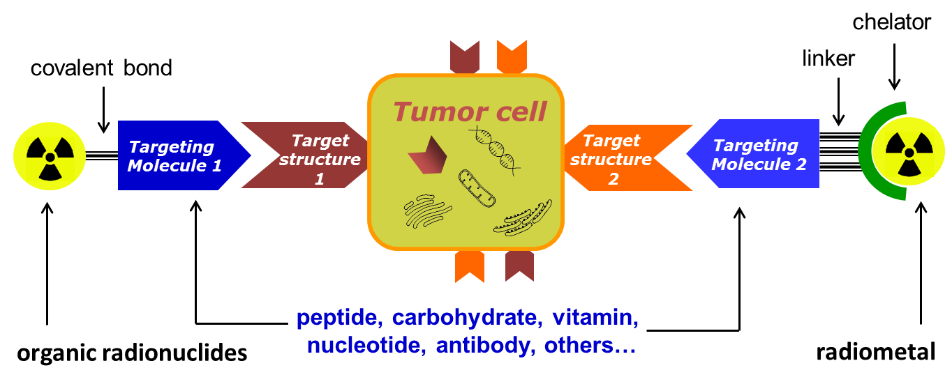
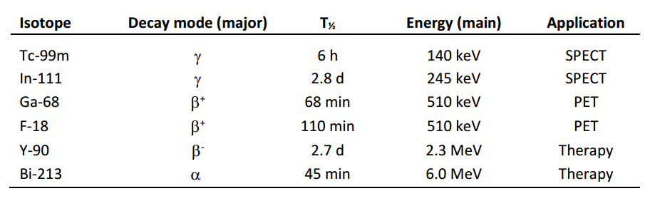
Radiopharmaceuticals of interest are conjugates that combine a radionuclide specific for a given application (Table 1) with a biological vector that is specific for a recognition element overexpressed by tumour cells (Figure 1). Depending on the application, the nature of the radionuclide and the vector molecule, different chemical and biological techniques are required for their preparation.

For preclinical evaluation, the radiopharmaceuticals are first tested in vitro (on cells) for their tumour-targeting properties, which usually includes experiments determining cellular uptake, affinity towards a given receptor/antigen, and other parameters (e.g., metabolic and enzymatic stability, hydrophilicity on the basis of physicochemical methods and new applied models, plasma protein binding, cell internalisation.). Promising candidates are then carried forwards to in vivo investigations (in mice) for determination of their pharmacokinetic and -dynamic properties employing biodistribution experiments and/or small animal imaging by µSPECT/PET/CT.

a. Figure 1: Schematic sketch of the general design of tumour targeting radiotracers.
b. Table 1: Examples of radionuclides for different applications in nuclear medicine.

Head of program line

Assoc.-Prof. Dr. Thomas L. Mindt

Head of Imaging Biomarkers

ta.ca.gbl.daibl@tdniM.samohT

Funded Project: Novel Multidentate Bifunctional Chelating Agents for the Development of Zirconium-89 Based Molecular Imaging Probes

Funding

Swiss National Science Foundation Grant N° 205321–157216

Principle Investigator

Prof. Dr. Thomas L. Mindt

Co-Investigator

Prof. Dr. Gilles Gasser (Chimie ParisTech)

Funding

kEUR 406

Duration

2018-2023

Zirconium-89 (89Zr) based radiotracers hold great promise as immuno-PET imaging agents in nuclear medicine (PET = positron emission tomography). However, insufficient stability of currently used radiometal complexes in vivo is a safety concern for clinical applications. We have developed a novel bifunctional, octadentate chelator (termed DFO*),[1] which provides 89Zr-labelled immuno-conjugates of remarkably improved stability in vitro and in vivo.[2] We are currently investigating further optimization of the new scaffold DFO* and its application for the development of a range of metal-based radiopharmaceuticals.

We have developed a novel bifunctional, octadentate chelator (termed DFO*),[1] which provides 89Zr-labelled immuno-conjugates of remarkably improved stability in vitro and in vivo.[2] We are currently investigating further optimization of the new scaffold DFO* and its application for the development of a range of metal-based radiopharmaceuticals.

[1] “An Octadentate Bifunctional Chelating Agent for the Development of Stable Zirconium-89 Based Molecular Imaging Probes” M. Patra, A. Bauman, C. Mari, C. A. Fischer, O. Blacque, D. Häussinger, G. Gasser, T. L. Mindt Chemical Communications 2014, 50, 11523-11525.

[2] “Comparison of the Octadentate Bifunctional Chelator DFO*-pPhe-NCS and the Clinically Used Hexadentate Bifunctional Chelator DFO-pPhe-NCS for 89Zr-Immuno-PET” D. J. Vugts, C. Klaver, C. Sewing, A. J. Poot, K. Adamzek, S. Huegli, C. Mari, I. E. Valverde, G. Gasser, T. L. Mindt, G.A.M.S. van Dongen European Journal of Nuclear Medicine and Molecular Imaging 2017, 44, 286-295.

[3] “A Solid Phase-Assisted Approach for the Facile Synthesis of a Highly Water Soluble Octadentate Zirconium-89 Chelator for Radiopharmaceutical Development” M. Briand, M. Aulsebrook, T. L. Mindt, G. Gasser, Dalton Transactions 2017, 46, 6387-1638

a. DFT optimized structure of Zr-DFO* (atom colour coding: white = carbon; blue = nitrogen; red = oxygen; magenta = zirconium); hydrogen atoms are omitted for clarity.

Funded Project: Metabolically Stabilized Peptidomimetics for Improved Tumour Targeting

Funding

Austrian Science Fund Grant N° P 31477-B28

Principle Investigator

Prof. Dr. Thomas L. Mindt

Co-Investigators

Prof. Markus Mitterhauser (LBIAD, Vienna), Dr. Wolfgang Kandioller (University of Vienna), Dr. Berthold Nock (Demokritos, Athens)

Funding

kEUR 430

Duration

2015-2020

Regulatory peptides are a class of biomolecules with ideal characteristics for the development of tumour-targeting radiopharmaceuticals. They exhibit a high, specific accumulation in tumours but not in healthy tissue and a favourable pharmacokinetic profile. A drawback of using such peptides for the selective delivery of attached radionuclides to tumours is their low stability due to rapid degradation by enzymes (proteases) before they can reach their target (tumours). It is known from the literature that enhancing the metabolic stability of a peptide carrier can substantially increase its uptake in tumours and metastases. Despite considerable research efforts directed towards the stabilization of the peptides without influencing their favourable biological characteristics, no general approach has yet been identified. We have recently introduced a novel “click chemistry” methodology to achieve this goal. We use metabolically stable 1,2,3-triazole heterocycles as biosiosteres of labile amide bonds of the peptides. We were able to show that the obtained, radiolabelled peptidomimetics exhibit an increased stability and, as a result, a significantly improved tumour uptake in mice.

[1] “1,2,3-Triazoles as Amide Bond Mimics: Triazole Scan Yields Protease-Resistant Peptidomimetics for Tumor Targeting” I. E. Valverde, A. Bauman,  C. A. Kluba, S. Vomstein, M. Walter, T. L. Mindt Angewandte Chemie International Edition, 2013, 52, 8957-8960.

[2] “1,2,3-Triazole Stabilized Neurotensin-Based Radiopeptidomimetics for Improved Tumor Targeting” A. Mascarin, I. E. Valverde, S. Vomstein, T. L. Mindt Bioconjugate Chemistry 2015; 26, 2143–2152.

a. The introduction of 1,2,3-triazoles as metabolically stable bioisosteres of amide bonds results in radiolabelled peptidomimetics with improved tumour-targeting properties.

Funded Project: Radiostar – Novel Chelators for Radio-Lanthanides and -Actinides

Funding

Austrian Research Promotion Agency, Bridge-1 Project

Principle Investigator

Prof. Dr. Thomas L. Mindt

Co-Investigators

Dr. Christoph Denk (Technical University of Vienna) Clemens Pichler (DSD-Pharma, AT)

Funding

kEUR 450

Duration

2020-2024

Radioactive labelled drugs (radiopharmaceuticals) are used in nuclear medicine for the diagnosis and therapy of cancer. In recent year, new therapeutic radiometals have emerged and showed great potential for targeted tumour therapy. For example, the alpha particle emitting radiometal actinium-225 (Ac-225) is particularly of current interest for endoradiotherapy. Medical applications of radioactive metals require chelators to attach them to tumour-targeting biomolecules (e.g., proteins). Such chelators must form complexes with the radiometal of sufficient stability so that the radioactive label does not dissociate from the bioconjugate in vivo. Suitable chelators are known for a number of radioactive transition metals. However, the size and complexation chemistry is different for the lanthanides and actinides (e.g., Ac-225). As a result, only few examples of chelators for these radiometals have been investigated and no ideal candidate has yet been reported. There is a clear need for new chelators that will facilitate and expedite clinical applications of the emerging and promising radioactive lanthanides and actinides.

Based on previously developed chemistry, novel chelators for lanthanides and actinides will be synthesized and attached to biomolecules. The bioconjugates will be labelled with radiometals and fully evaluated in vitro (e.g. on cells) for their stability and tumour targeting properties (e.g., affinity towards the target). Promising candidates will be selected and used for studies in mice bearing tumour xenografts (biodistribution experiments and small animal PET/CT imaging).

Funded Project: Marker für die Aktivität von Albumin-bindenden Verbindungen

Funding

Austrian Science Fund No P 32886-B

Principle Investigator

Prof. Dr. Petra Heffeter (Medical University of Vienna)

Co-Investigators

Prof. Dr. Thomas Mindt (LBIAD), Prof. Dr. Christian Kowol (University of Vienna)

Funding

kEUR 400

Duration

2020-2024

Es gibt mehr und mehr Anhaltspunkte, dass sich Krebszellen in ihren Strategien Nährstoffe aufzunehmen von gesundem Gewebe unterscheiden. Speziell ihre erhöhte Aufnahme von Plasmaproteinen, wie zum Beispiel Albumin, stellt einen vielversprechenden Ansatzpunkt für die Therapie dar. Dies konnte bereits durch den klinischen Erfolg von Medikamenten wie Abraxane (ein Albumin-Paclitaxel Nanopartikel) oder Aldoxorubicin (ein Albumin-bindendes Doxorubicin) unterstrichen wird. Auf Grund dessen ist es von großem Interesse weitere Medikamente zu entwickeln, welche über Albumin spezifisch in die Krebszelle transportiert werden. Zu diesem Zweck haben wir kürzlich die erste Albumin-bindende Prodrug von Oxaliplatin entwickelt. Diese neue Verbindung, welche vielversprechende Aktivität gegen Dickdarmkrebs zeigt, wäre eine gute Möglichkeit das häufig in Kolorektalkrebs verabreichte FOLFOX Schema (Folinsäure, 5-Fluorouracil und Oxaliplatin) zu verbessern. Doch obwohl das Potential der Albumin-vermittelten Therapie für die Behandlung von Krebserkrankungen schon klinisch geprüft wurde, ist das Wissen über die Unter-schiede im Albumin Metabolismus zwischen Krebszellen und gesunden Zellen überraschend gering.

Die Ziele des hier präsentierten Antrags sind einerseits das bessere Verstehen der Albumin Homöostase in Dickdarmkarzinomen und andererseits das Verwenden dieses Wissen für die präklinische Entwicklung einer neuen Albumin-bindenden Oxaliplatin-Prodrug.

Ein Schwerpunkt bildet die Rolle potenzieller Biomarker (z.B. K-RAS) in der Albuminaufnahme von malignen Zellen. Dies soll im Rahmen dieses Projektes durch verschiedenen zell-und molekularbiologischer Methoden analysiert werden. Zusätzlich wird eine neue PET Bildgebungsplattform, unter der Verwendung von 89Zr-markierten Albuminkonjugaten, etabliert. Diese soll die Untersuchung der Albuminaufnahme in Tumorknoten in vivo in einer nicht invasiven und quantitativen Art und Weise ermöglichen und so als Hilfsmittel für die Stratifizierung zukünftiger Patientenkohorten herangezogen werden können.

a. a. Different suggested pathways of the cellular uptake of albumin and therefore albumin-drug/prodrug conjugates.

Bi-Metallic Complexes for Synergistic and Theranostic Applications

Funding

Austrian Science Fund (AT), Agence Nationale de la Recherche (FR) No I 5721

Principle Investigators

Prof. Dr. Thomas Mindt (LBIAD, University of Vienna; AT). Prof. Dr. Gilles Gasser (Chimie ParisTech; FR)

Funding

kEUR 450

Duration

2021-2025

The use of metals in medicine is very versatile, ranging from different diagnostic to therapeutic applications. This project aims at the combination of two metal-based therapies in order to increase the therapeutic efficacy in comparison to the individual treatments alone. Namely, we will combine photodynamic therapy (PDT) with nuclear radioendotherapy (RET). For PDT, metal-containing compounds are excited by light and subsequently, because in aqueous biological media, reactive oxygen species are produced that destroy the surrounding diseased tissue (tumors, infections (viruses) etc.). RET utilizes radioactive metals whose ionizing radiation is toxic and also destroys the surrounding diseased tissue. Goal of this project is to chemically combine two suitable metals (ruthenium and rhenium) in one construct that allows for simultaneous PDT and RET. The bi-metallic compounds will be conjugated to a targeted biomolecule (e.g., antibody) that serves as carrier for the selective transport of the therapeutic cargo to the diseased site while sparing healthy tissue. The bi-metallic compounds and conjugates thereof will be tested biologically (e.g., on cells) in order to investigate the interplay of the two therapies.